Select language : EN | BH
Call Icon
Call us at
Gleneagles Hospital
Call Icon
Call us at
Mount Elizabeth Orchard
Gleneagles Hospital
Klatskin Cancer Surgery

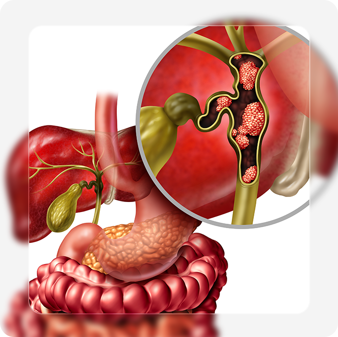
Klatskin tumours are a type of bile duct cancer, known as ‘cholangiocarcinoma’. This is a form of malignant cancer that tends to develop in the central biliary region (including the biliary ducts). While Klatskin tumours are not large in size, they can exhibit an aggressive biologic behaviour by causing an obstruction in the intrahepatic biliary ducts and jaundice.

Due to the location of these tumours, patients tend to develop symptoms at advanced stages and the tumours can be difficult to resect. However, this may differ from case to case, as some patients may have jaundice in the early stages, which may compel them to seek early medical help.

Ideally, complete resection of the Klatskin tumor (whenever feasible) should be performed, as this is associated with better long-term survival. For certain patients, liver transplant can be an option for treatment in specialised centres. Overall, the prognosis of Klatskin tumours is poor, but new surgical and endoscopic treatments allow better disease control.

Epidemiology of Klatskin tumours

Generally, Klatskin tumours are rare. However, it is observed that Asians are more prone to develop these tumours, especially males and elderly patients above the age group of 50 years. Also, Klatskin tumours comprise about 25% of cholangiocarcinomas. The remaining cholangiocarcinomas can occur in the liver or the distal bile duct.

Klatskin Tumour Staging

The Klatskin tumour staging can be determined by using a well-known staging system, Bismuth-Corlette system.

The Bismuth-Corlette system classifies Klatskin tumours into 4 main categories based on the location of these tumours. These include:

  • Type 1 Klatskin tumours: These tumours exist in the common hepatic duct, but are far away from the location where the hepatic ducts meet.
  • Type 2 Klatskin tumours: Such tumours mainly develop in the common hepatic duct where both the left and right hepatic ducts meet.
  • Type 3 Klatskin tumours: The staging of Klatskin tumour is classified as type 3, when the tumours are located in one of the hepatic ducts.
  • Type 4 Klatskin tumours: This is the most advanced, in which the tumours are detected in both the left and right hepatic ducts.

Klatskin Tumour Diagnosis

Radiographic tests are required to Diagnose and assess Klatskin tumours. Ideally, histological or tissue confirmation by endoscopic, radiologically-guided biopsy or surgical biopsy can be performed. Often times, histological confirmation may not always be feasible.

  1. MRI/MRCP – MRCP will help determine the location of Klatskin tumours, and determine resectability (surgical planning)
  2. Percutaneous transhepatic cholangiography (PTC) – this is a radiological procedure in which an external tube is inserted to drain bile and helps delineate the bile duct anatomy for surgical planning
  3. Endoscopic retrograde cholangiopancreatography (ERCP) – this can help place an internal biliary stent to drain bile and obtain tumour tissue for biopsy

Do consult a hepatobiliary surgeon once diagnosed with Klatskin’s tumour. About 30-40% of such tumours are potentially resectable, and patients with successfully resected tumours have better overall survival.

Treatment for hepatobiliary conditions,such as those affecting the liver and gallbladder,are complex and require specialized expertise to manage effectively.

For a personalized assessment, contact our clinic at 6737 8878 or drop us a message here.
Your welfare is our priority and we will be in touch with you soon.

Top